CT scan: the right test when speed and anatomic detail both matter
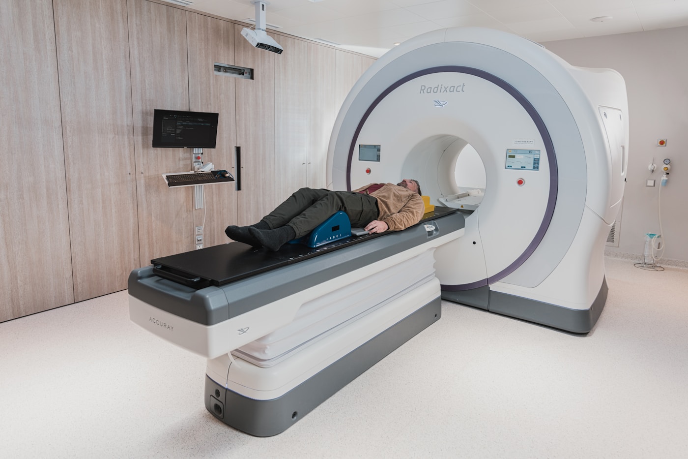
A CT scan uses a rotating X-ray beam and a computer to build thin cross-section pictures — and, when needed, a 3-D view — of bone, lung, organs, and blood vessels. The scan itself is usually measured in seconds to a few minutes.
This is the practical guide we use in clinic. CT is often the best first imaging test for sudden pain, trauma, suspected kidney stones, many chest problems, and questions that a plain X-ray cannot settle.
Head & Neck
Head trauma, suspected bleed, sudden worst headache
Fast, excellent for acute blood, skull fracture, and mass effect. Time matters.
MRI preferred later for many non-emergent brain questions.
Sinus disease when medical therapy fails or complications suspected
CT sinus maps bone and air cells for ENT planning; low-dose protocols exist.
Uncomplicated colds and first-line sinusitis do not need imaging.
Facial trauma, suspected mandible/orbit fracture
CT shows complex facial bones better than plain films.
Isolated simple nasal injury may start with exam ± limited films.
Chest
Abnormal chest X-ray (nodule, mass, unexplained infiltrate)
Chest CT characterizes nodules, lymph nodes, and parenchyma X-ray cannot resolve.
A first cough in an otherwise well patient starts with exam ± chest X-ray.
Suspected pulmonary embolism (when criteria met)
CT pulmonary angiogram (CTPA) is the workhorse test for PE in most adults.
Use validated PE pretest tools. V/Q or other paths if contrast/kidneys prohibit CTPA.
Staging or follow-up of known chest malignancy
CT chest (± abdomen/pelvis) is a backbone of many staging pathways.
PET/CT or MRI may be added by the oncology team for specific questions.
Abdomen & Pelvis
Suspected appendicitis, diverticulitis, or acute abdomen in adults
CT abdomen/pelvis is the most complete single study for these common emergencies.
In children and pregnant patients, ultrasound (and sometimes MRI) is preferred first.
Suspected kidney stone (flank pain, hematuria)
Non-contrast CT is the most sensitive test for ureteral stones.
Ultrasound first in pregnancy and often in young/recurrent-stone patients to limit dose.
Unexplained weight loss, suspected occult mass, cancer staging
Contrast-enhanced CT surveys organs, nodes, and peritoneum efficiently.
Endoscopy still owns the inner lining of GI tract; imaging complements, not replaces.
Spine, Bone & MSK
Complex fracture (pelvis, tibial plateau, calcaneus, spine bony detail)
CT is the reference standard for complex bone anatomy and surgical planning.
Simple suspected fracture often starts with X-ray. MRI if the question is cord, disc, or ligament.
Hardware, fusion, or postoperative bony detail
CT handles metal better than MRI for bone and hardware position.
Soft-tissue infection or cord compression after surgery may still need MRI.
Occult fracture when X-ray is negative but suspicion stays high
CT can confirm bony injury X-ray missed (e.g., some scaphoid, pelvic, spine questions).
MRI is often better for occult hip fracture in older adults and for bone-marrow edema.
Knowing when not to CT is as important as knowing when to order it.
Pregnancy — avoid ionizing radiation unless the study is essential and no substitute exists. Tell us if you are or may be pregnant.
Children — start with the lowest-dose path that answers the question (often ultrasound or limited X-ray).
Soft-tissue sports injury (meniscus, rotator cuff, ligament) — map usually needs MRI at another facility; we start with exam, X-ray, and ultrasound when those answer the question.
Most chronic low-back pain without red flags — imaging often does not change first-line care. Red flags need urgent evaluation.
Gallbladder / pelvic / obstetric questions — ultrasound first in most cases.
Thyroid nodules — ultrasound, not CT, is the dedicated test.
Known severe iodinated-contrast allergy or unstable kidney function — we may choose a non-contrast protocol, premedication pathway, or a different modality.
Contrast — plain-language rules
Contrast is chosen because the clinical question needs it, not automatically.
CT head for acute bleed / trauma
Usually without IV
Blood and bone are already bright; contrast can hide acute hemorrhage.
CT abdomen/pelvis for pain, infection, mass, cancer follow-up
Often IV ± oral
Organs and vessels need enhancement; oral contrast outlines bowel when ordered.
CT kidney stone protocol
Without IV, no oral
Stones are dense; contrast can hide them.
CTPA (lungs for clot)
IV only, timed arterial
Need the pulmonary arteries opacified.
CT sinus (routine)
Usually without IV
Air and bone; contrast reserved for complicated infection/tumor.
Before IV contrast we review allergy history and, when indicated, recent kidney function (creatinine / eGFR). Metformin is usually held around the time of iodinated contrast per current protocol. Breastfeeding can generally continue after iodinated CT contrast.
What to expect day of the scan
Check in a few minutes early with photo ID, insurance card, and order if referred.
Change into a gown if clothing has metal zippers, underwire, or embellishments in the scan field.
Tell the technologist about implants, prior contrast reactions, asthma, kidney disease, diabetes/metformin, and possible pregnancy.
You lie still on the table. You may be asked to hold your breath for a few seconds. The machine is open — not a long closed tube.
IV contrast can cause a brief warm flush and a metallic taste. That is expected. Trouble breathing, hives, or swelling — say so immediately.
Total room time is usually 15–30 minutes depending on the protocol.
You can typically resume normal activity. Drink extra water after IV contrast unless on a fluid restriction.
Radiation — honest language
CT uses ionizing radiation. Dose varies by body part, patient size, and protocol. A clinically indicated CT carries a very small added lifetime risk that is generally far smaller than the risk of missing the diagnosis the scan was ordered to find.
We use the principle of ALARA (as low as reasonably achievable): the right study, the right protocol, no extra phases that do not change care.
We do not sell "whole-body screening CT" to well people. If you have had multiple CTs this year, tell us — your clinician will weigh cumulative exposure.
CT Exam Menu
Common studies we perform. Your clinician's order specifies the exact protocol.
Ready to schedule?
Call us or message through the contact form. Established patients can request imaging through this office.